Please feel free to use our e-mail back service.
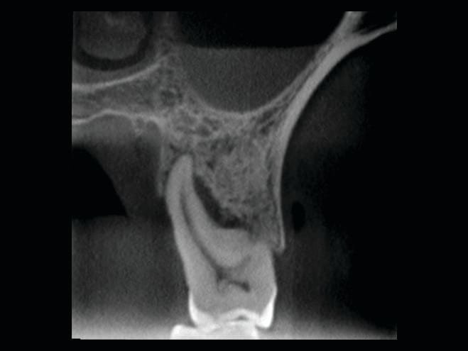
The patient reported tooth sensitivity in the left maxillary second molar. A small volume cone beam CT of the left posterior maxilla was acquired with the Veraviewepocs 3D R100. The sagittal and coronal views showed severe vertical bone loss associated with the palatal root of the left maxillary second molar, along with mucosal thickening in the left maxillary sinus.


