Please feel free to use our e-mail back service.
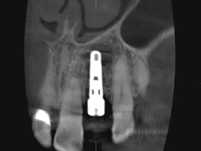
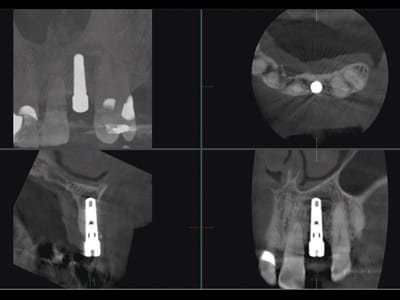
The patient was seen for a routine follow-up visit following implant placement in the area of the left maxillary lateral incisor. The implant had been placed 3 months earlier. The coronal, sagittal, and axial planes revealed a large, round, well defined, non-corticated, low density area associated with the apical aspect of the implant. The high resolution images also shows absence of the buccal cortical plate confirming a poor prognosis for the case due to peri-implantitis.