Please fill out this contact form. We will come back to you soon.
Exposure parameters: 100 kV – 5 mA – 9,4 s – 560 mGycm2, FOV: 10 x 8 cm
Clinical information and questions
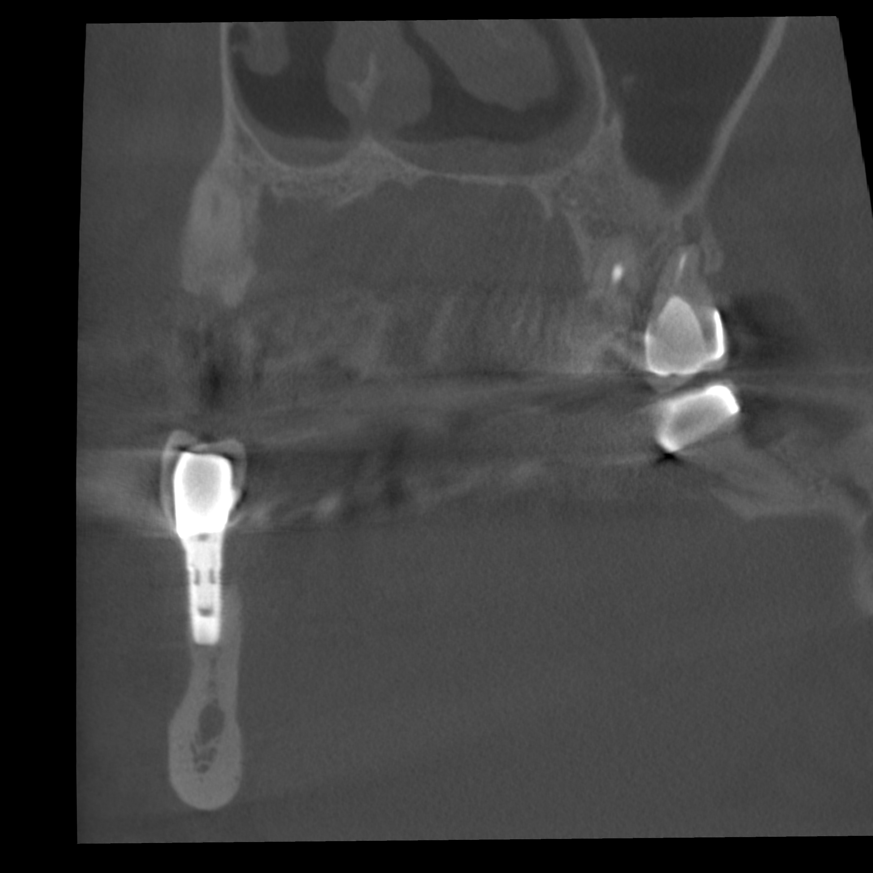
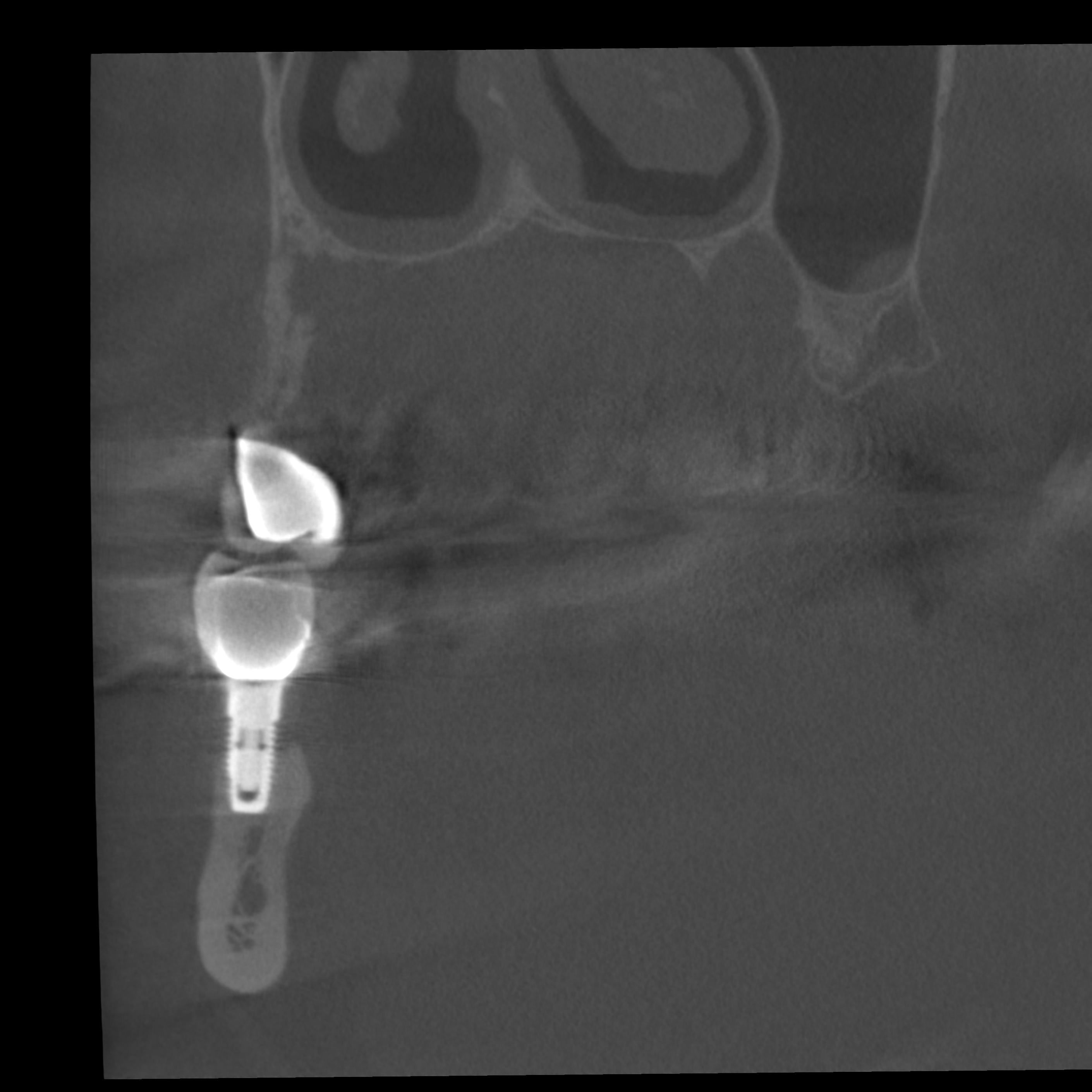
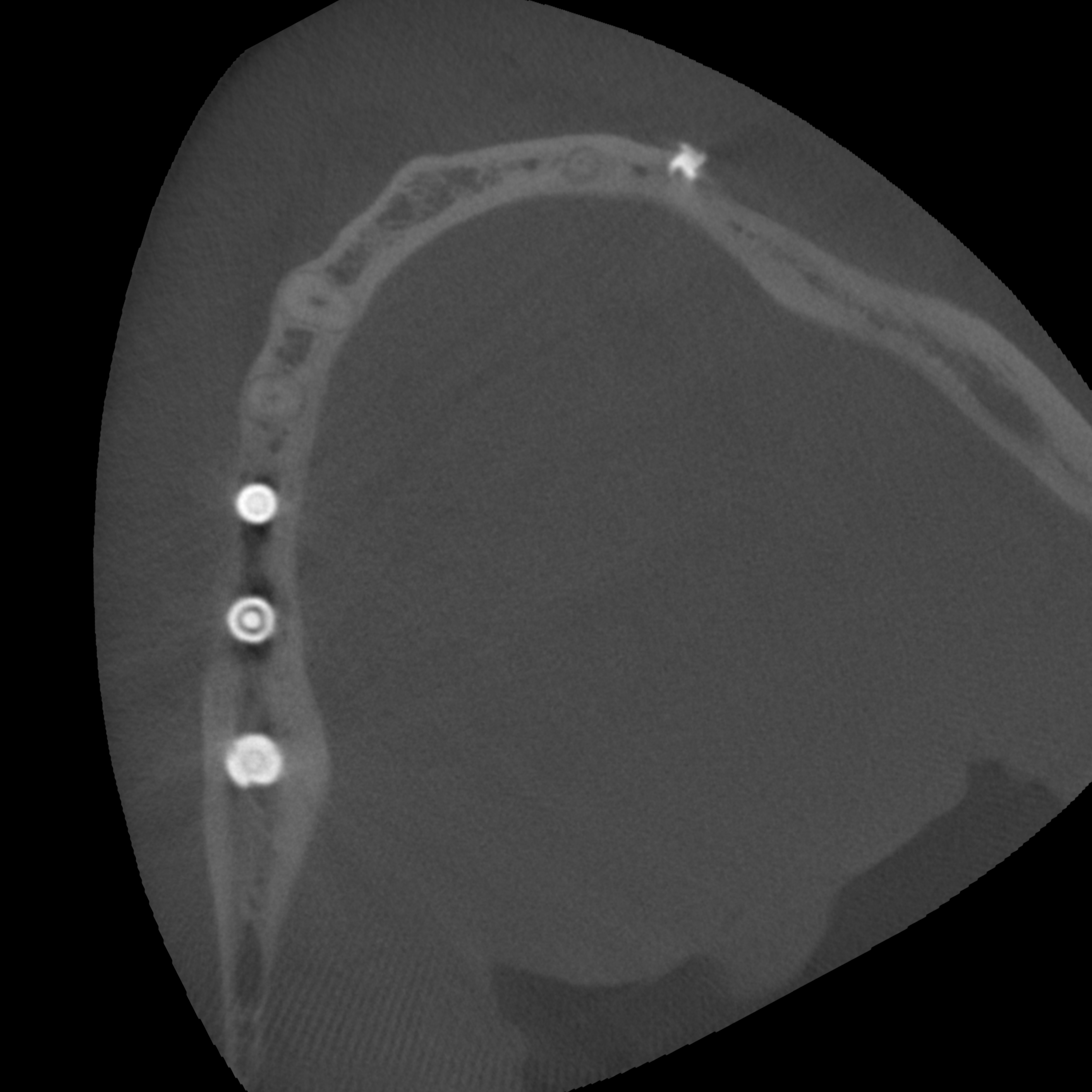
The CBCT examination was performed to visualize anatomically important neighboring structures and to quantitatively record the bone supply and exclude osseous lesions prior to the insertion of implants in the upper and lower jaw.
Description
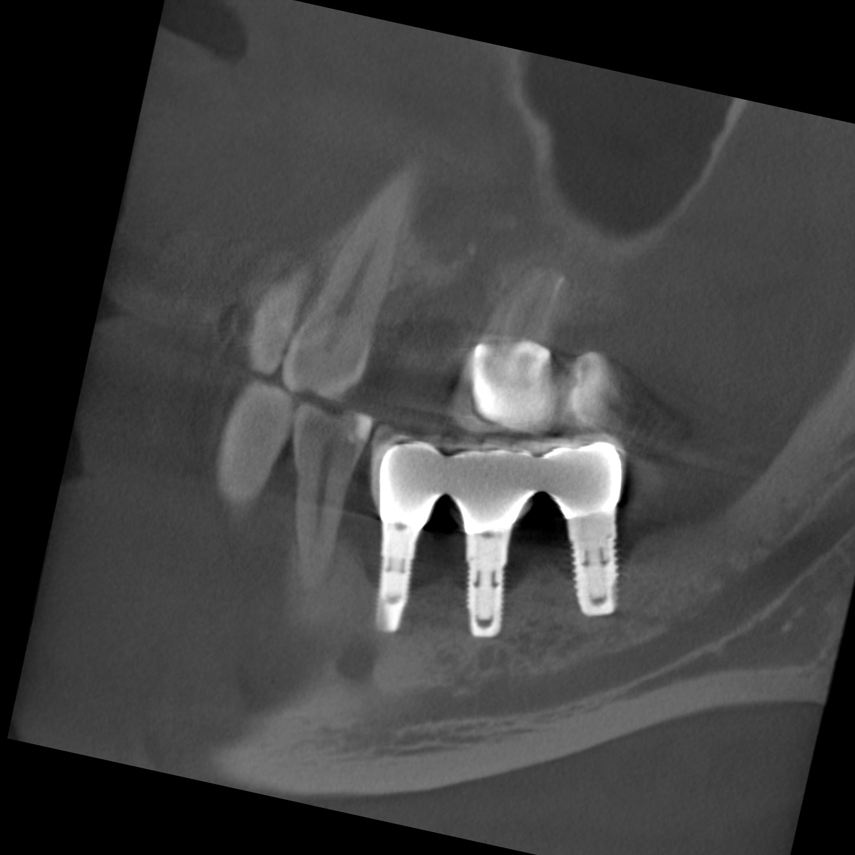
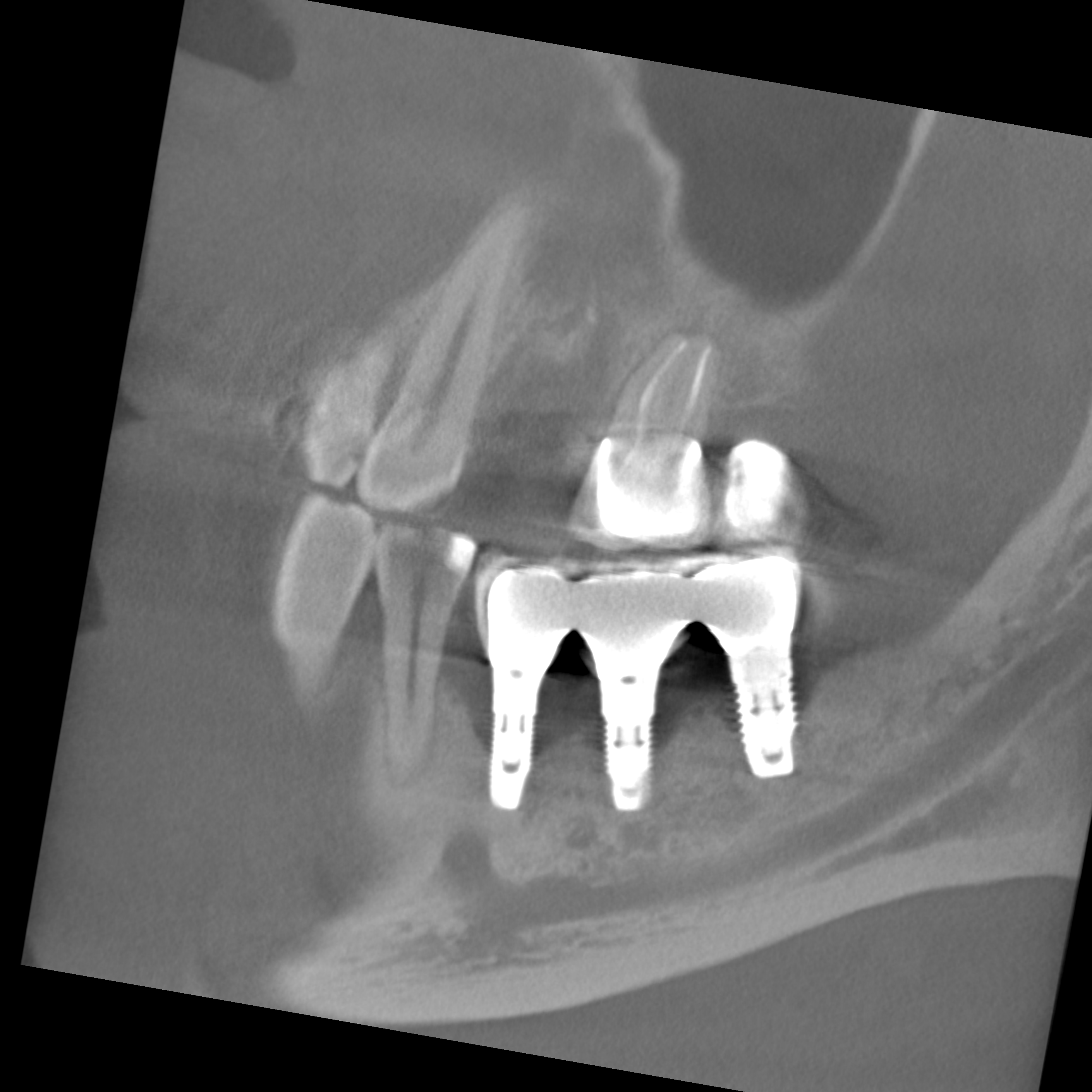
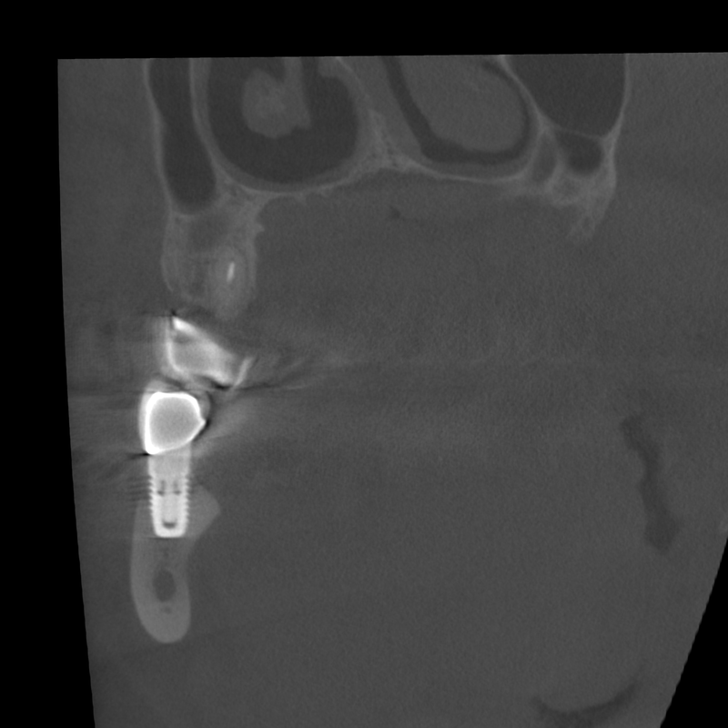
Although partial extinction artifacts are shown at the height of the implant shoulder and the prosthetic restoration, the implants in regions 45-47 show consistently pronounced, funnel-shaped osteolyses. The locoregional cancellous bone structure shows clear sclerosis.
Findings
This finding is certainly compatible with periimplantitis of the implants in region 45-47, at the same time there is a clear reactive sclerosis of the cancellous bone in the region mentioned.


