Please fill out this contact form. We will come back to you soon.
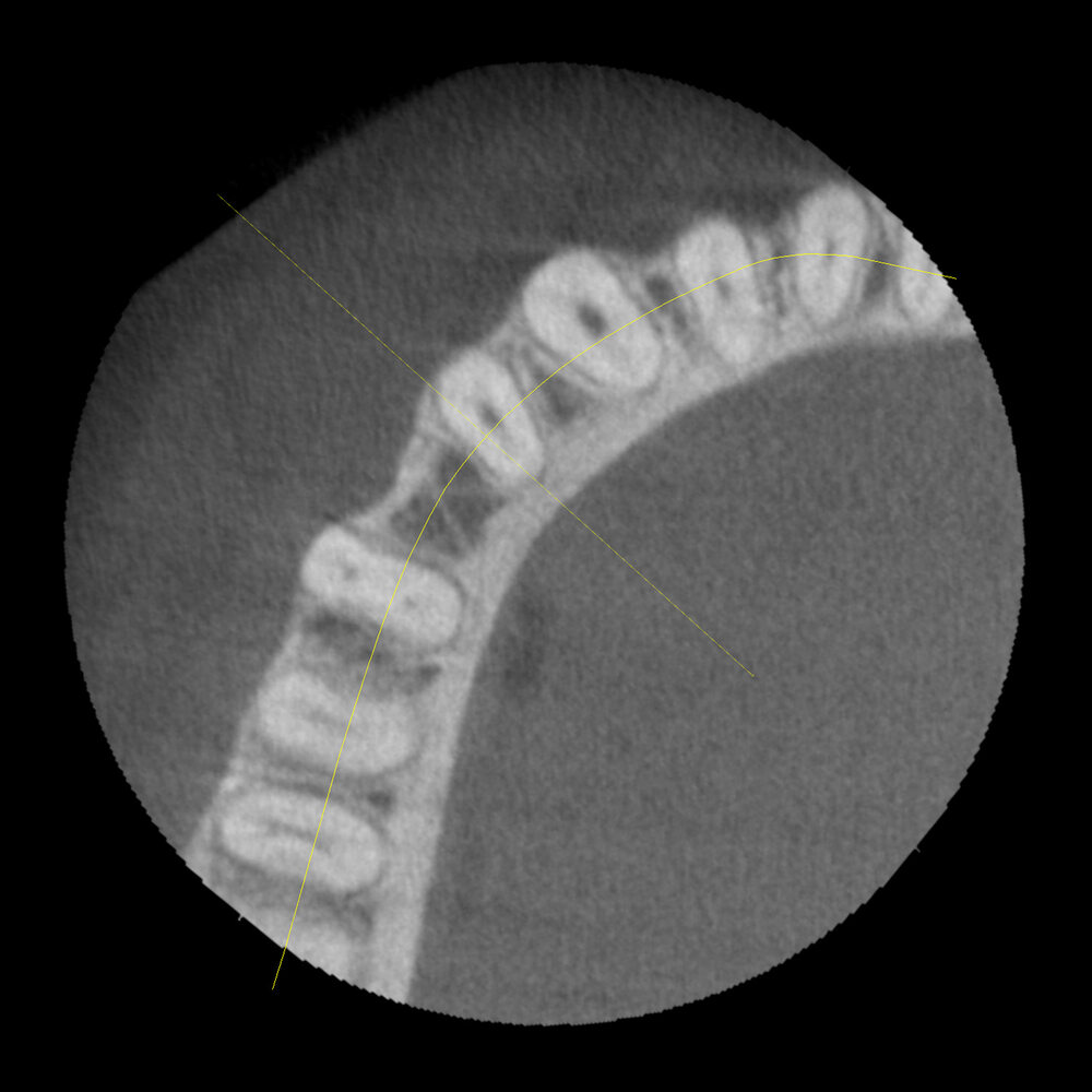
Exposure parameters: 100 kV – 7 mA – 9,4 s – 304 mGycm2, FOV: 4 x 4 cm
Clinical background and issue
In this particular case, the layered panoramic image showed a hyperdense structure projected onto the root tip of tooth 44. A repeat DVT investigation was performed with a view to clarifying the spatial extent and morphology of the change.
Description
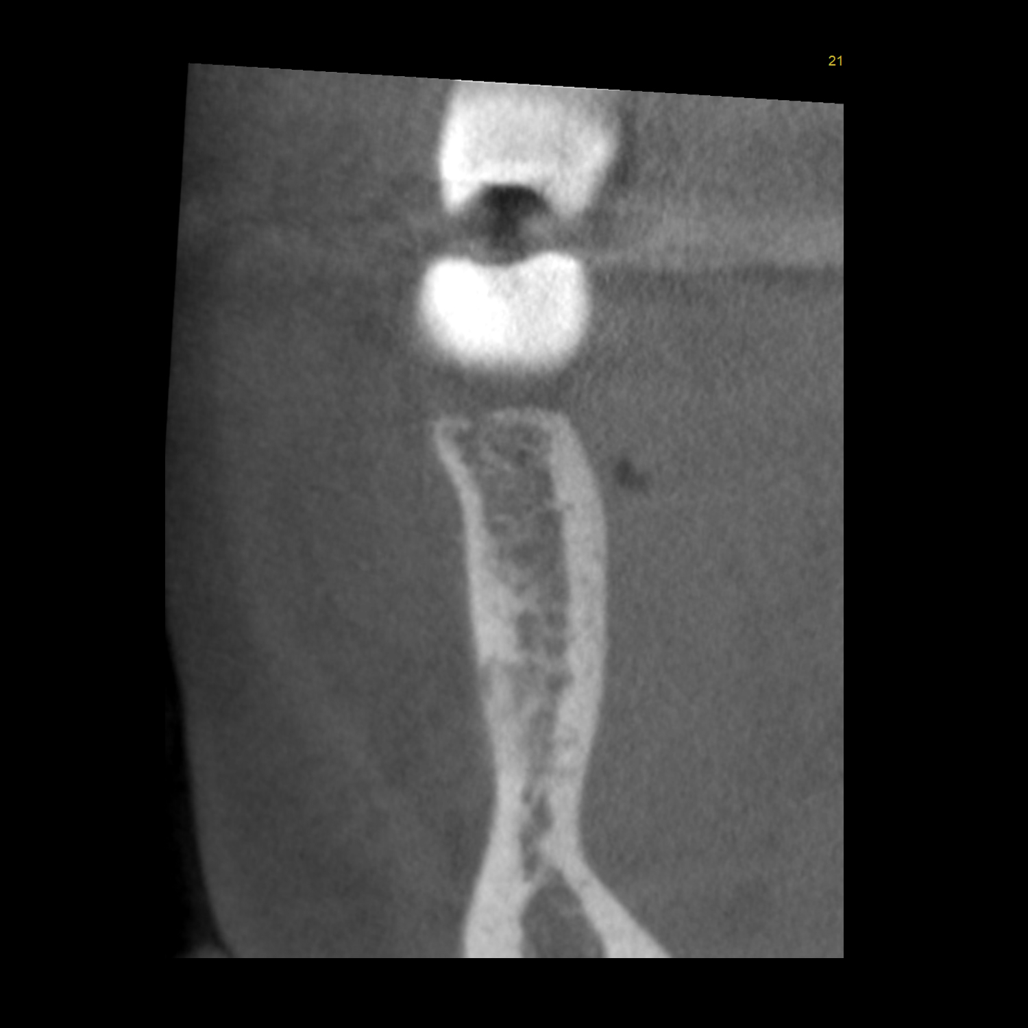
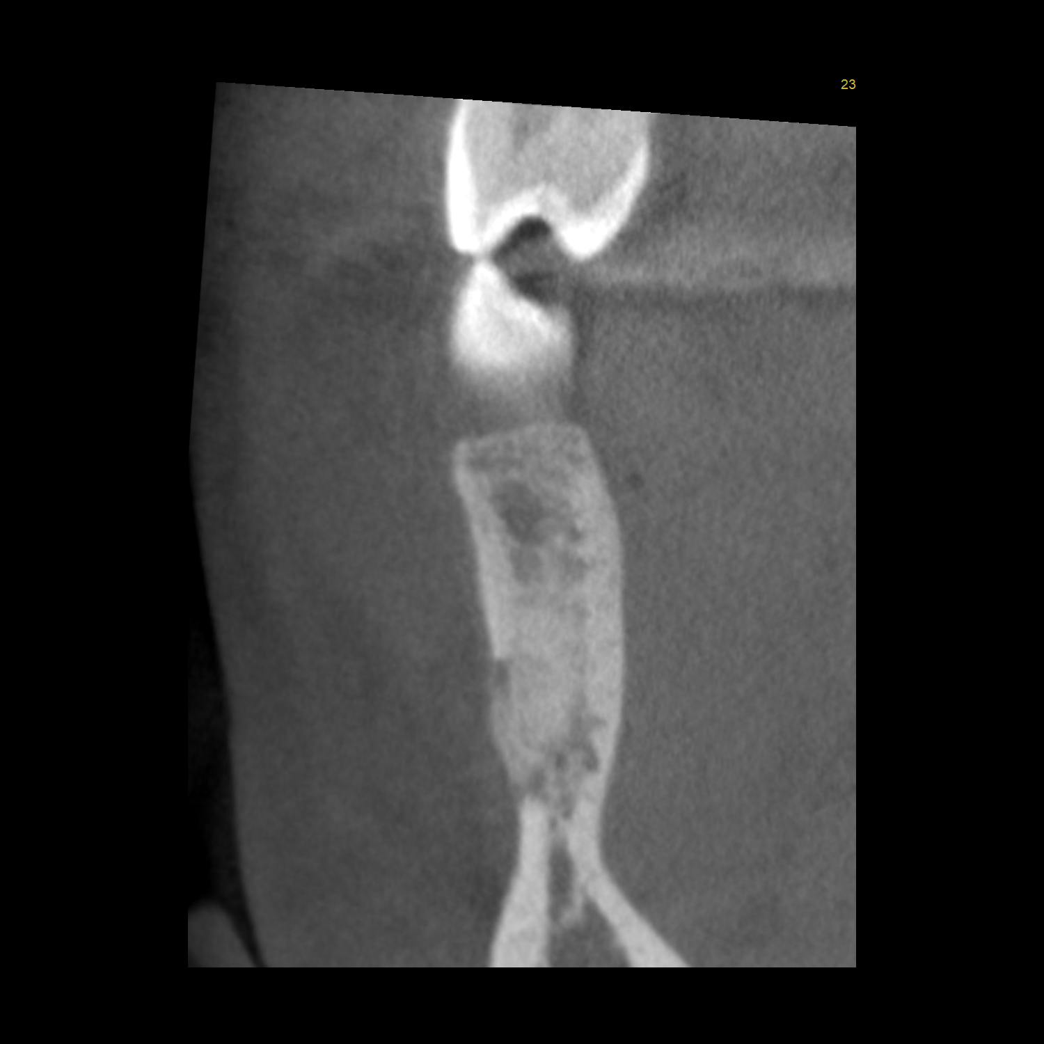
At the periapical area of tooth 44, a sharply defined, round, heterogeneous, and largely hyperdense structure can be seen extending as far as the compact bone in the buccal direction and crowding this out completely.
Findings
These findings are certainly compatible with periapical stage II-III cemental dysplasia and should be monitored over time. For the time being, we recommend the patient should be seen again in a year.


