Please feel free to use our e-mail back service.
Case
CBCT analysis of a patient referred for dental implant treatment planning of an extended edentulous space in the posterior mandible with two missing premolars. Due to the reduced vertical and horizontal dimensions of the alveolar ridge and the location of the mental foramen, safe dental implant insertion is only possible in the region of the first premolar. The desired future position and angulation of the implant can be evaluated in three dimensions demonstrating the need for simultaneous guided bone regeneration procedures at the crestal region and the distances to the mental foramen and incisive canal.
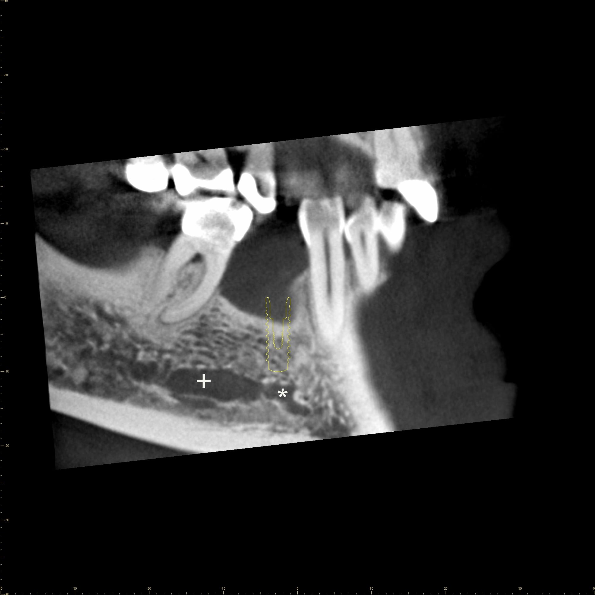
Figure 1A: Sagittal CBCT image with mandibular (+) and incisive canal (*)
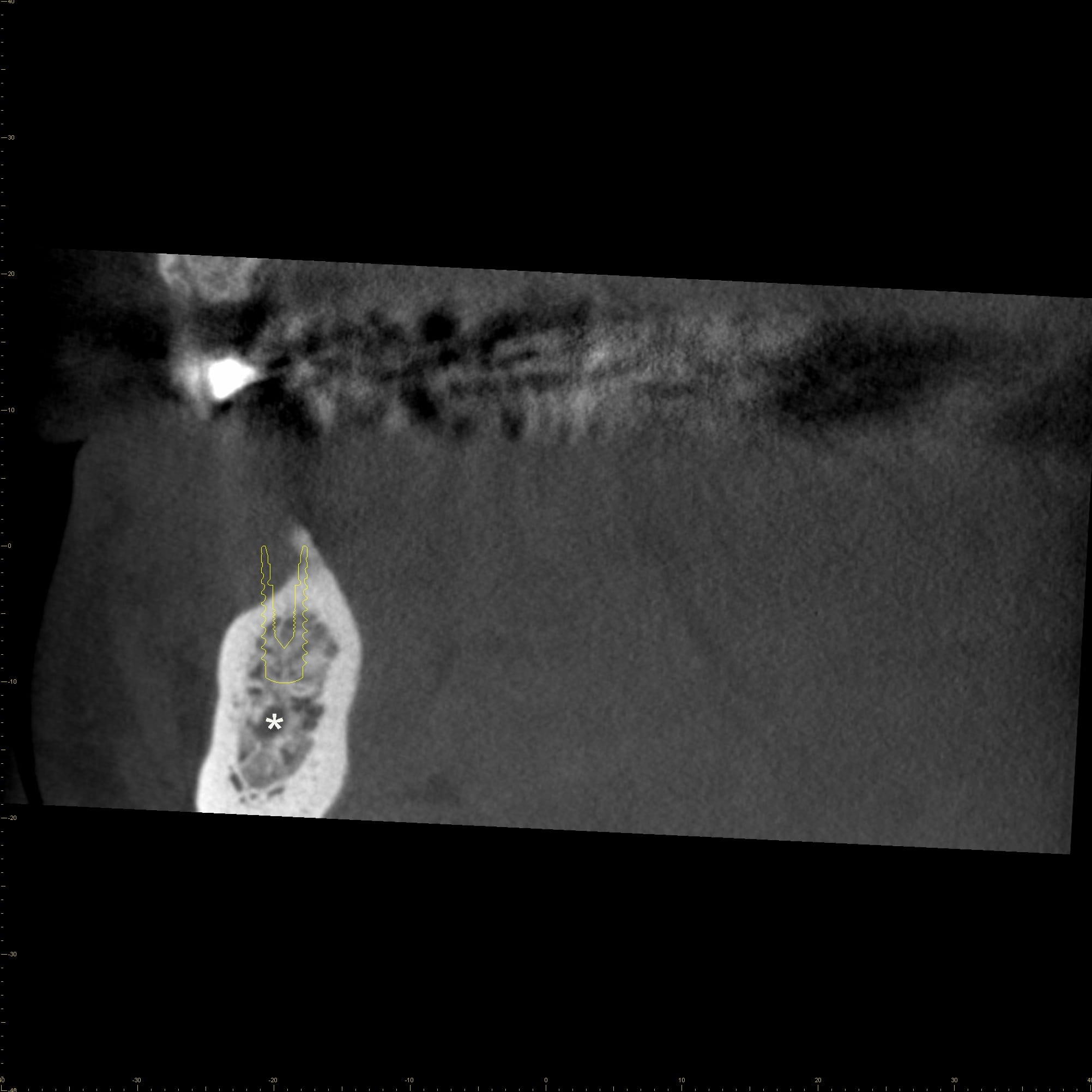
Figure 1B: Coronal CBCT image with incisive canal (*)
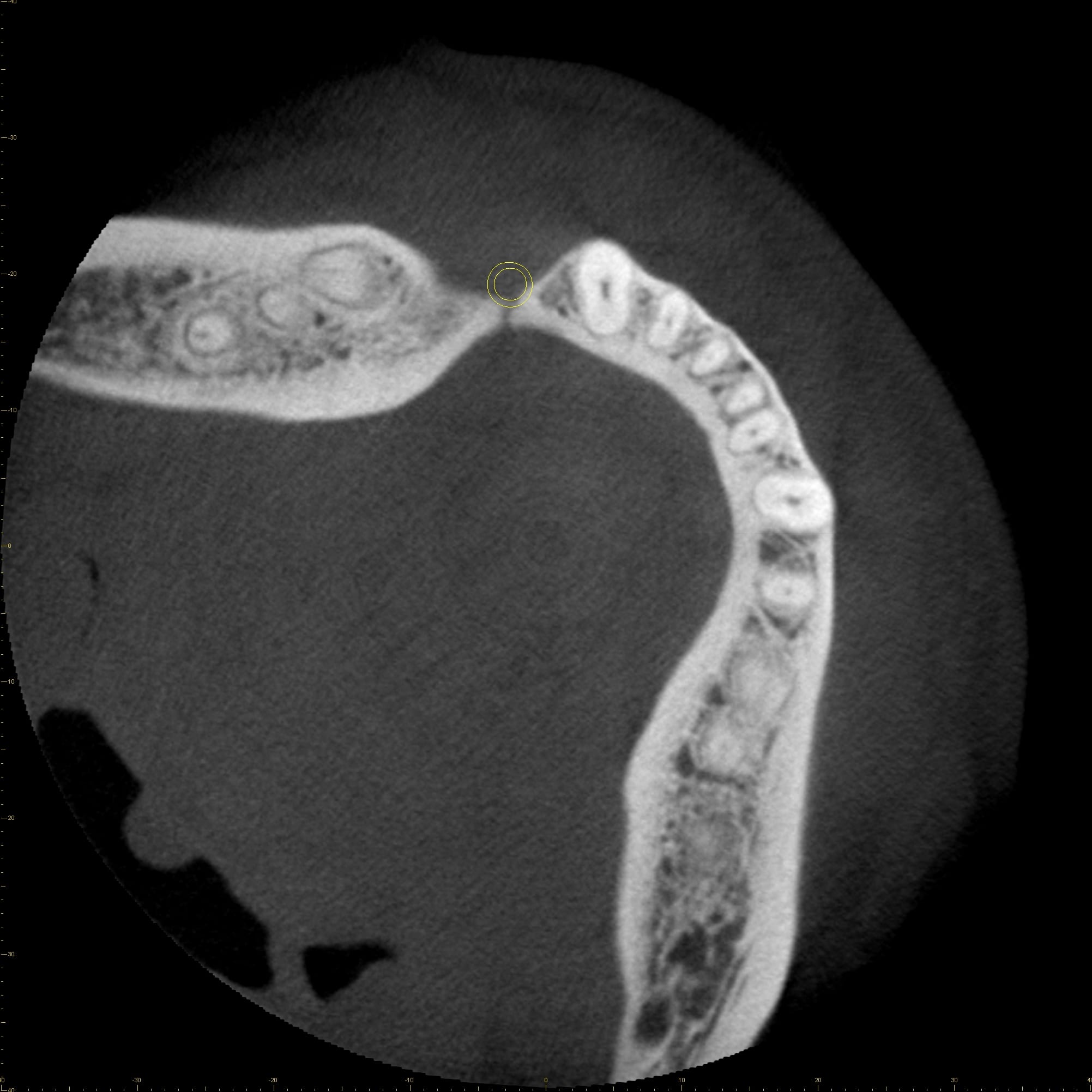
Figure 1C: Axial CBCT image
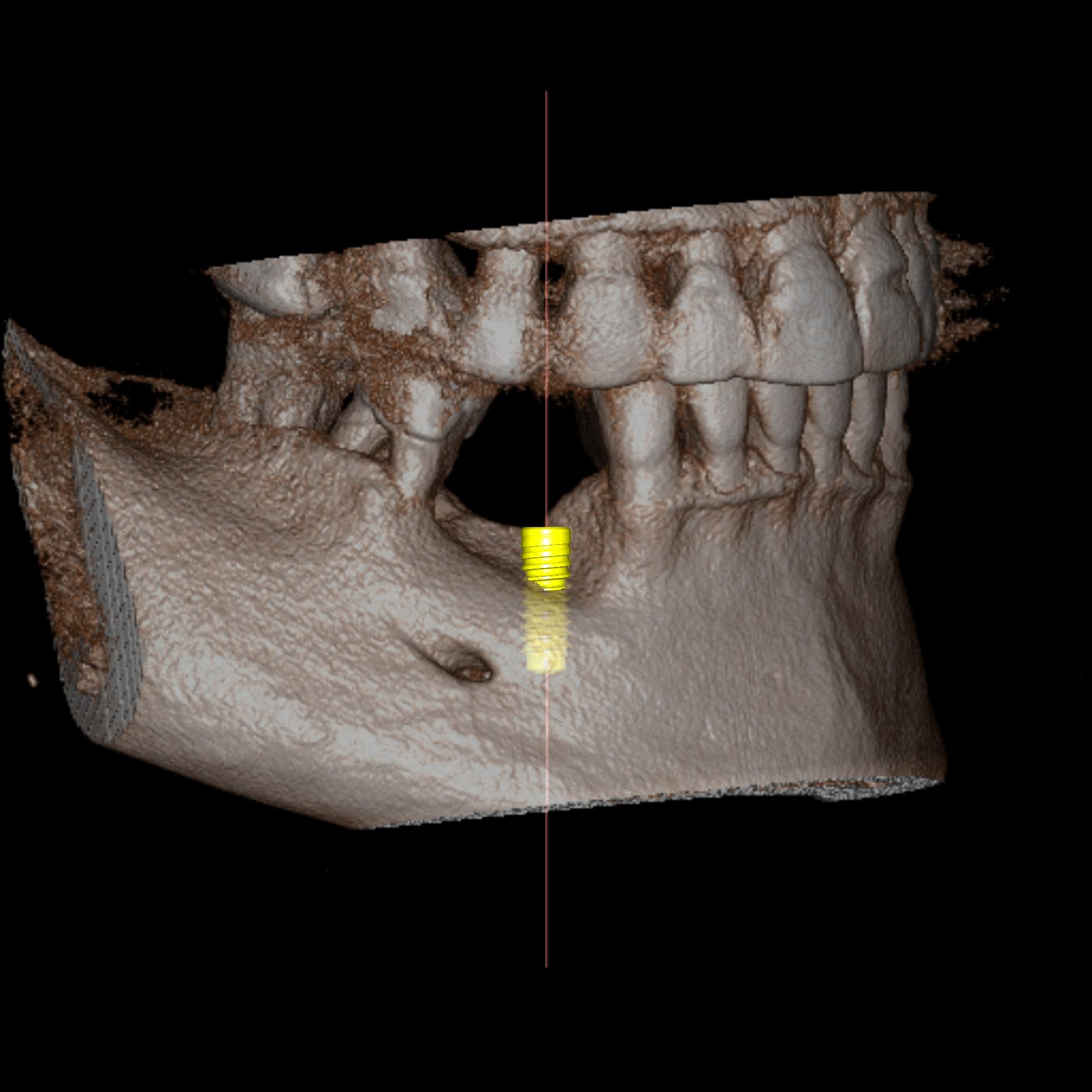
Figure 1D: Volume rendered data set
Figure 2
CBCT analysis further reveals furcation involvement of the first molar (Figures A to C) and a root remnant of the first premolar located distally to the apex of the canine (Figures D to F).
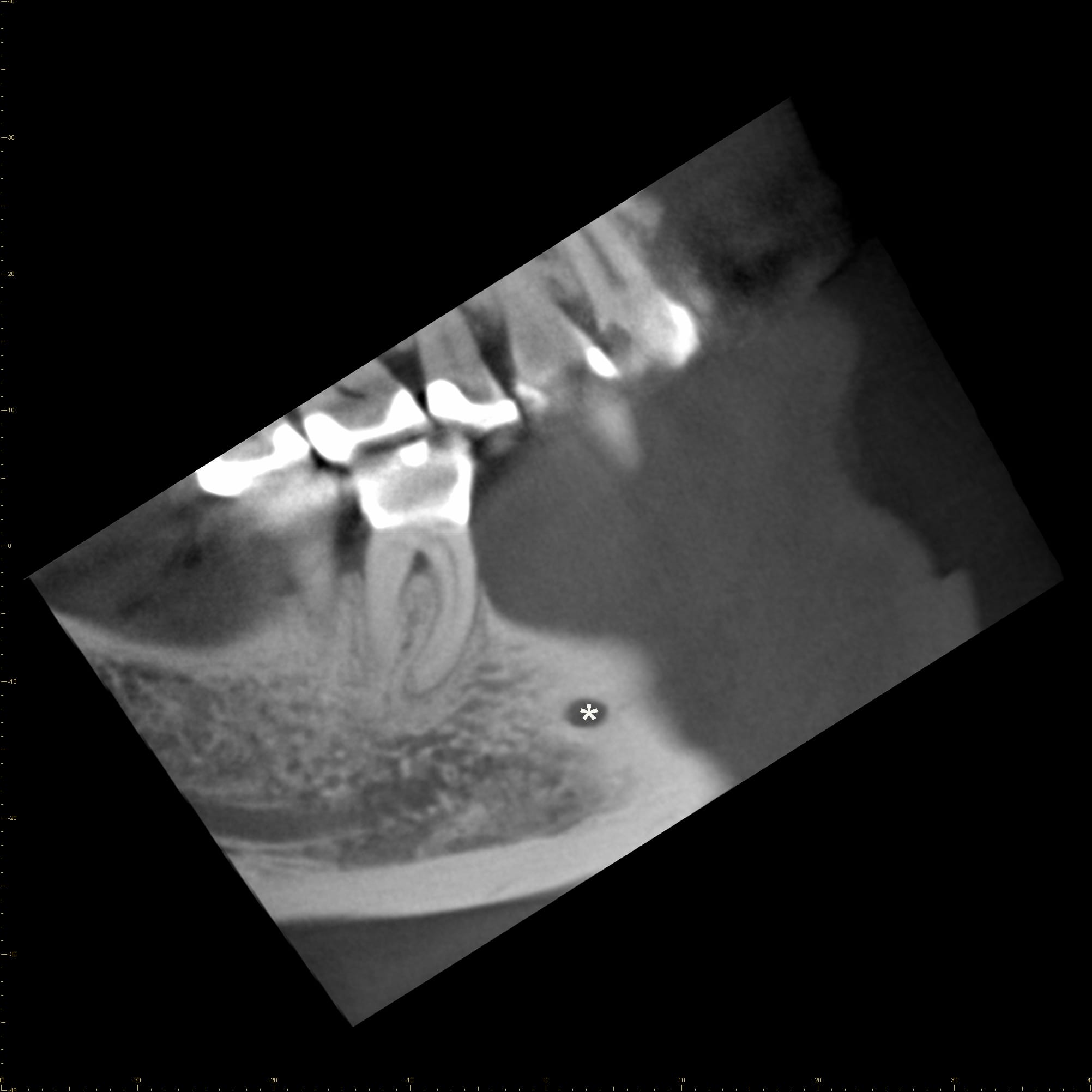
Figure 2A: Sagittal CBCT image with mental foramen (*)

Figure 2B: Coronal CBCT image with mandibular canal (*)
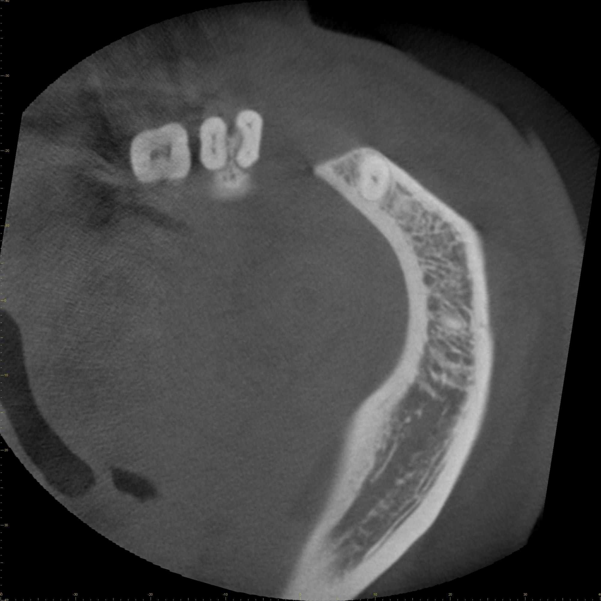
Figure 2C: Axial CBCT image
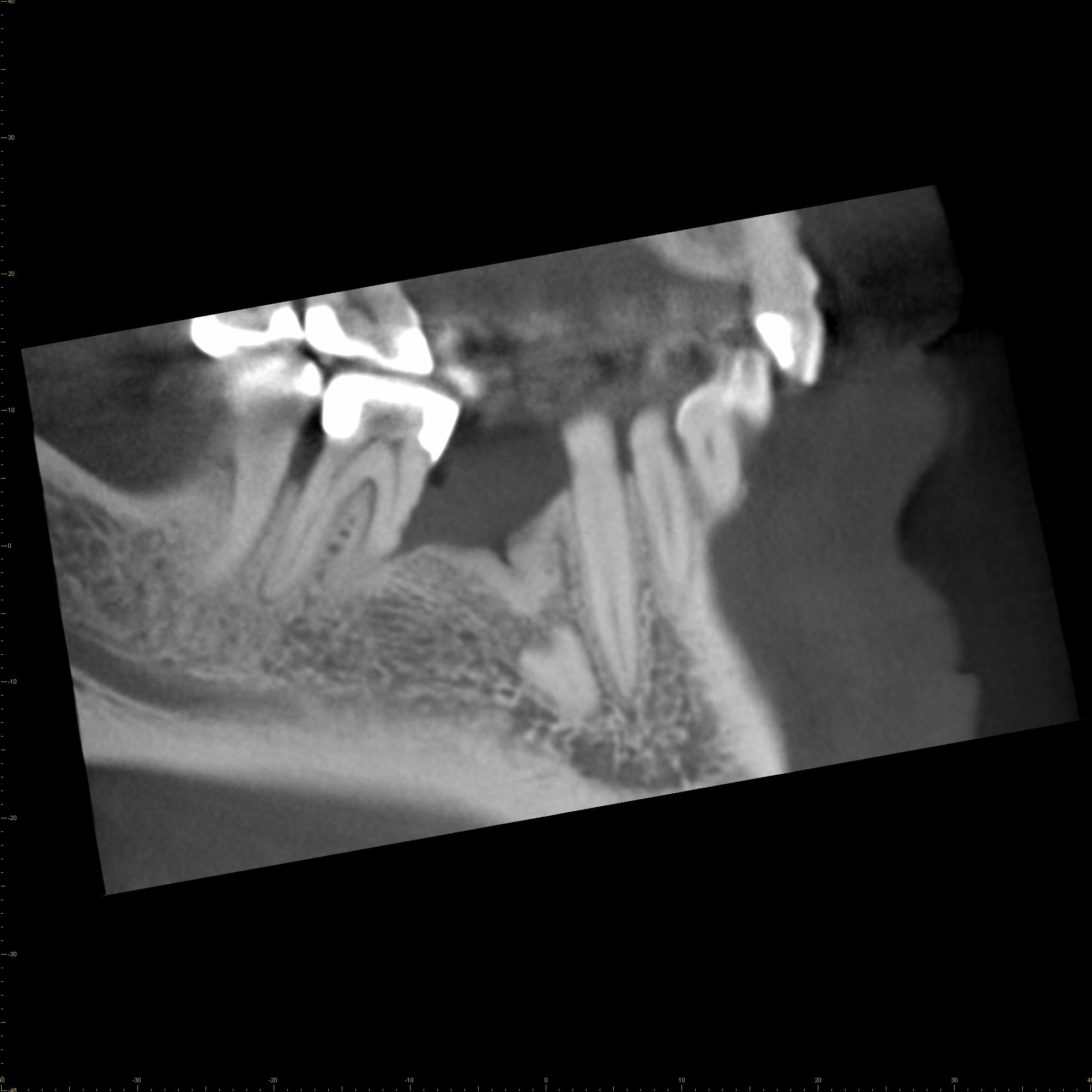
Figure 2D: Sagittal CBCT image
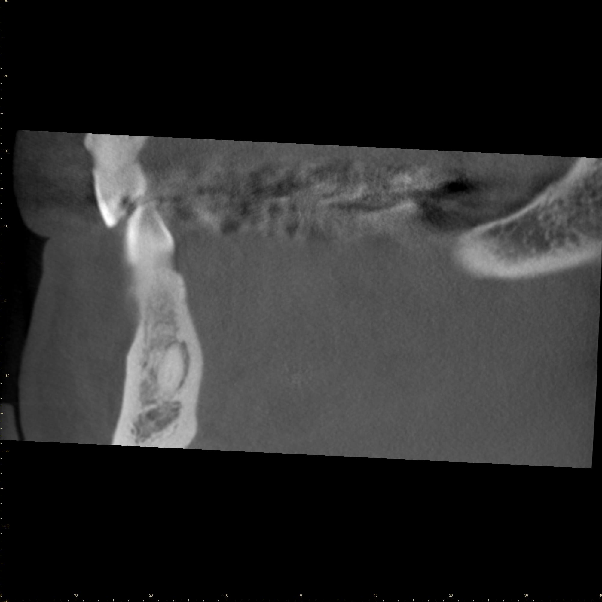
Figure 2E: Coronal CBCT image
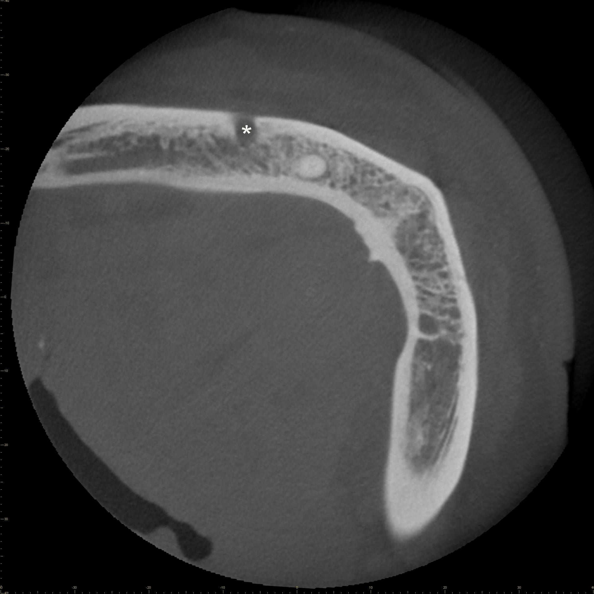
Figure 2F: Axial CBCT image with mental foramen (*)


