Nutzen Sie unseren E-Mail-Service.
Case: Patient Male, 73 years old
Figure 1
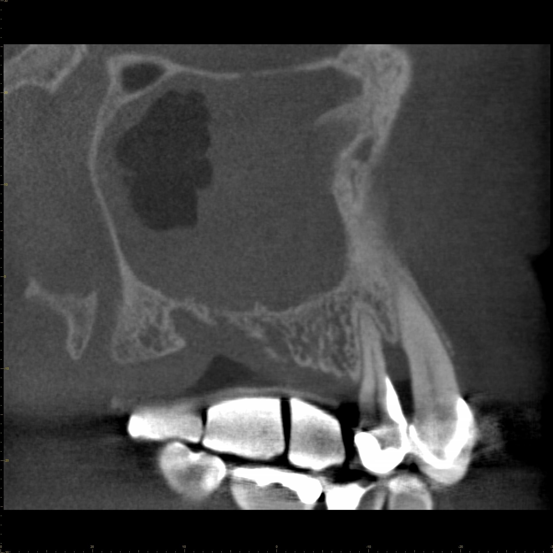
A 73-year old male patient was referred for dental implant treatment planning in the right posterior maxilla. Upon initial examination he did not report any acute medical problems. He had lost the molars and the second premolar due to periodontal and endodontic complications. CBCT analysis of the right posterior maxilla using of a radiographic template (small FOV: 6 x 5 cm) exhibits reduced vertical bone height in the molar area, bone loss between the canine and the first premolar, and opacification of the right maxillary sinus.
The tentative diagnosis was a maxillary sinusitis of rhinogenic origin. Based on these findings, the patient was referred to the ear, nose, throat (ENT) specialist for further diagnosis and treatment.
Figure 1B
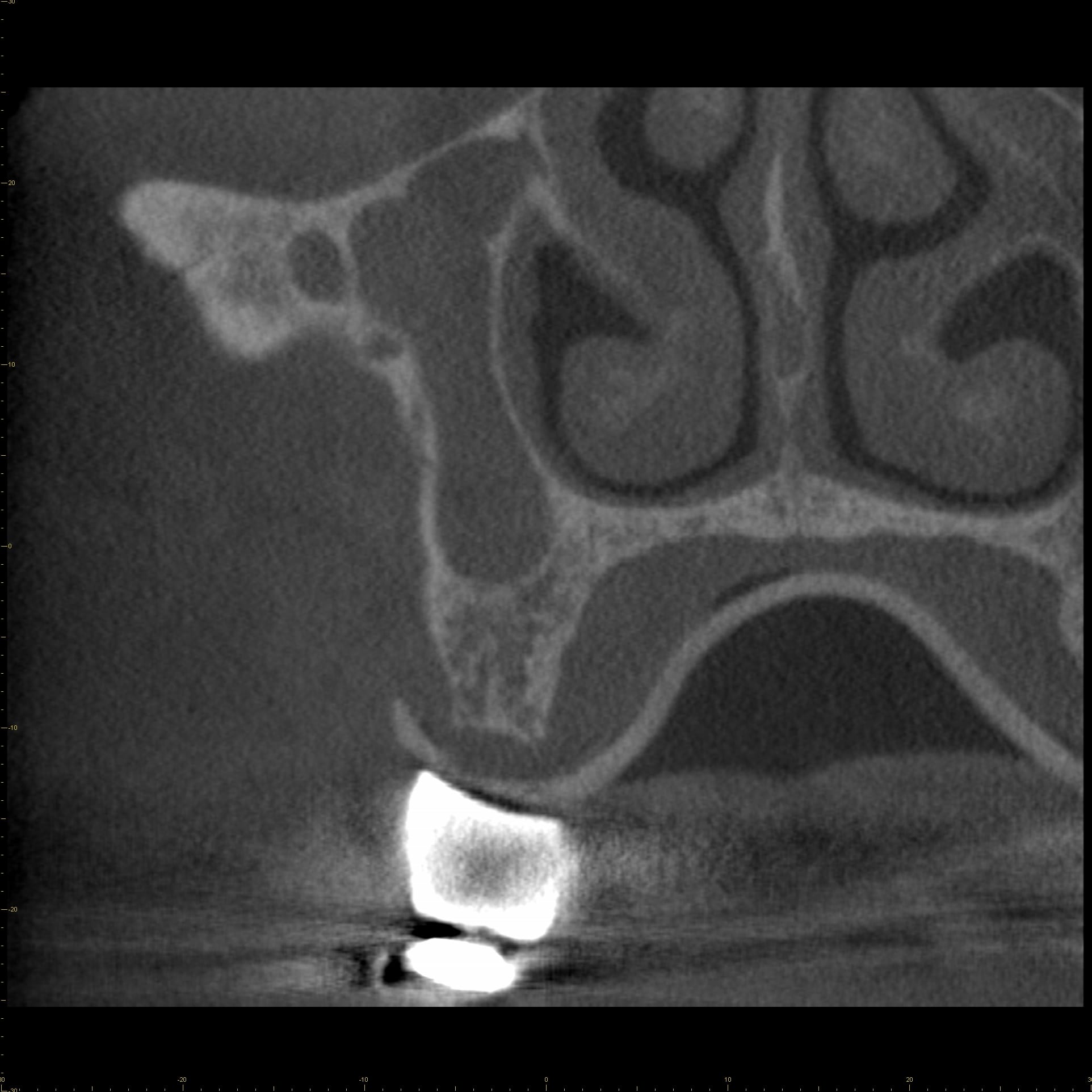
Coronal CBCT image, second premolar region
Figure 1C
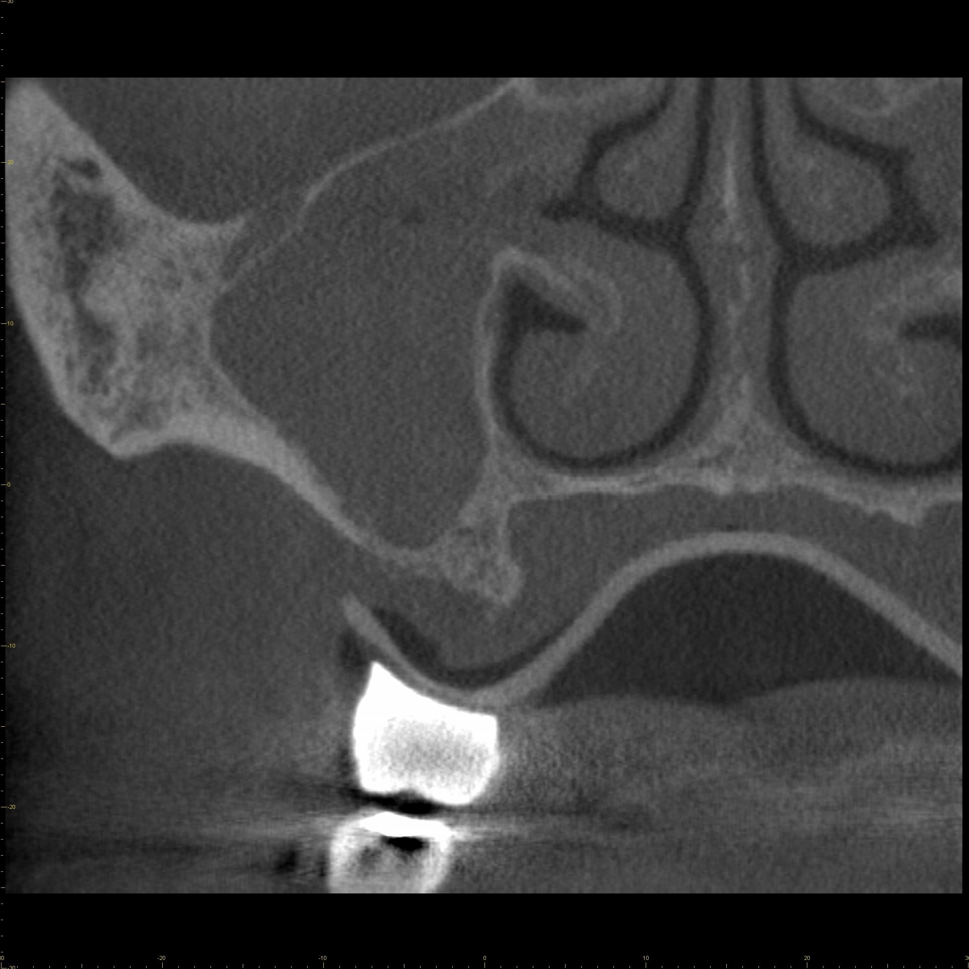
Coronal CBCT image, first molar region
Figure 1D
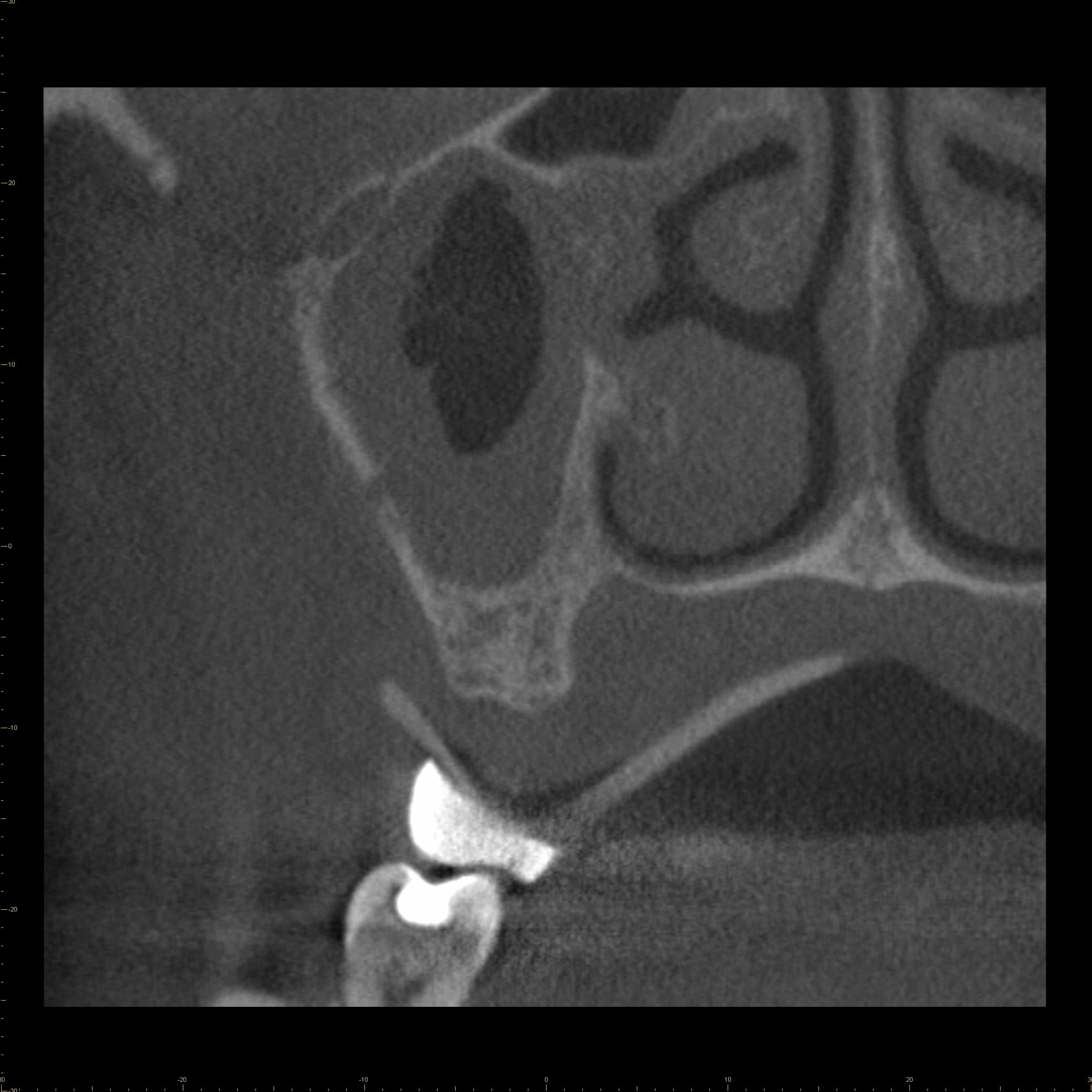
Coronal CBCT image, second molar region
Figure 1E
Axial CBCT image, bone loss between canine and first premolar is visible


