Nutzen Sie unseren E-Mail-Service.
Case:
Patient Male, 41 years old
Figure 1
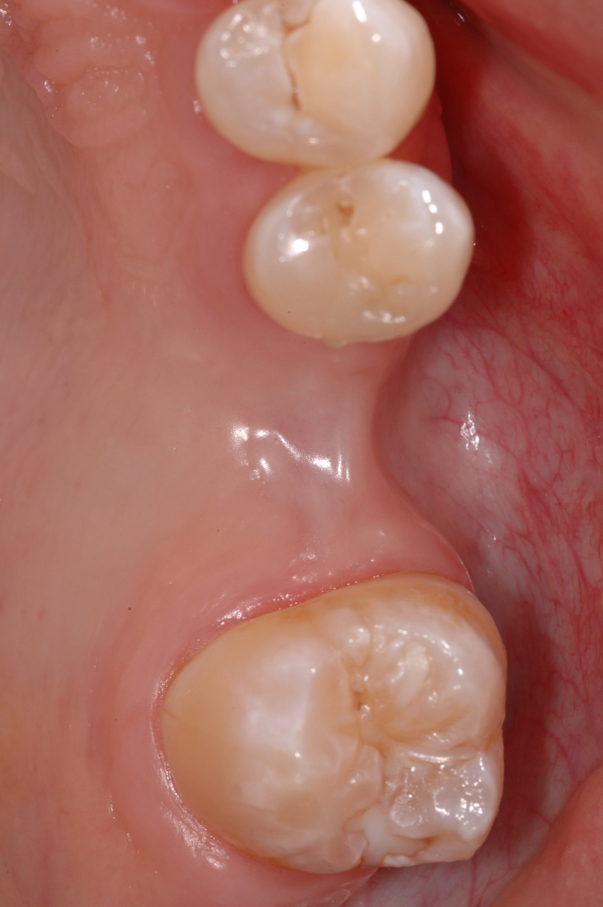
Initial clinical presentation of a 41-year old male patient referred for dental implant treatment planning. The patient had an extraction of the first left maxillary molar following a failed root canal treatment one year ago.
Figure 2
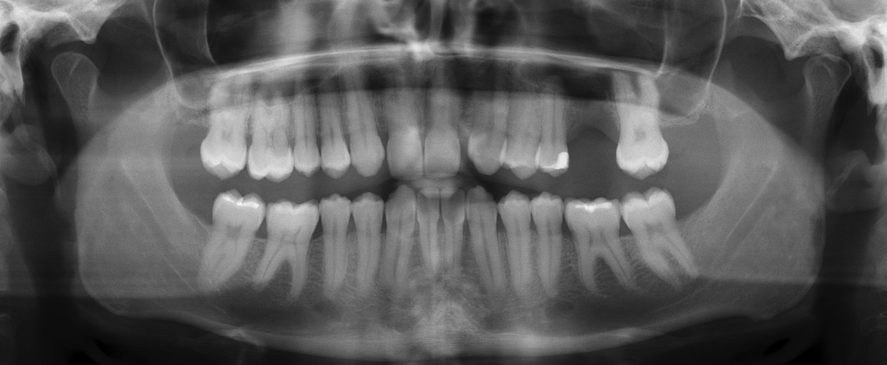
Panoramic view of the patient exhibiting the single-tooth gap in the upper left posterior maxilla. Additional findings include agenesis of both lateral maxillary incisors. Otherwise, there are no pathologic findings in the radiograph.
Figure 3
CBCT analysis of the left posterior maxilla including the missing first molar area (small FOV: 6 x 5 cm): the evaluation of the images exhibits well pneumatized maxillary sinus without any pathologic findings, premolars and a second molar without fillings or other signs of decay, and a reduced vertical bone height and even missing bone towards the maxillary sinus in the region of the first molar.
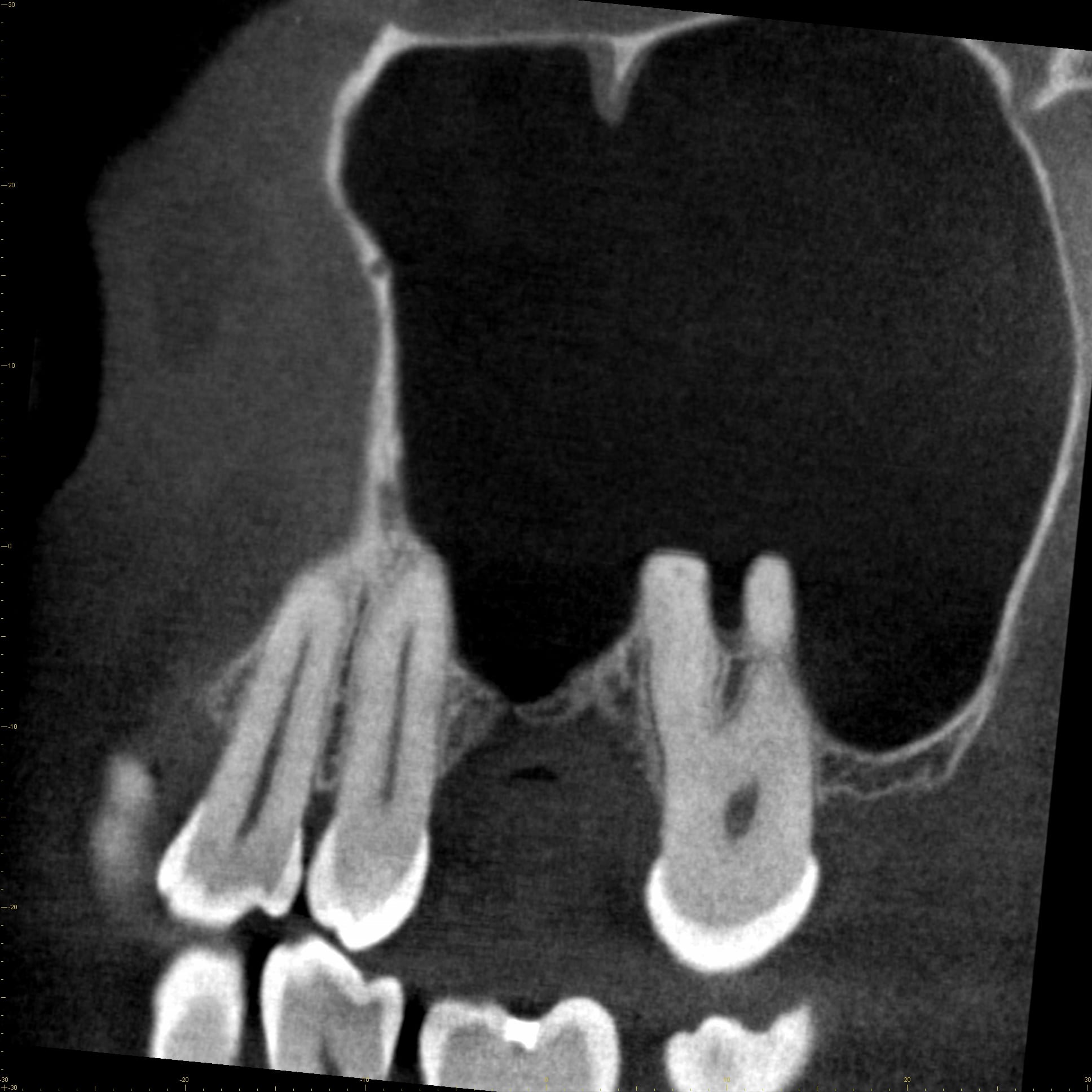
Figure 3A: Sagittal CBCT image
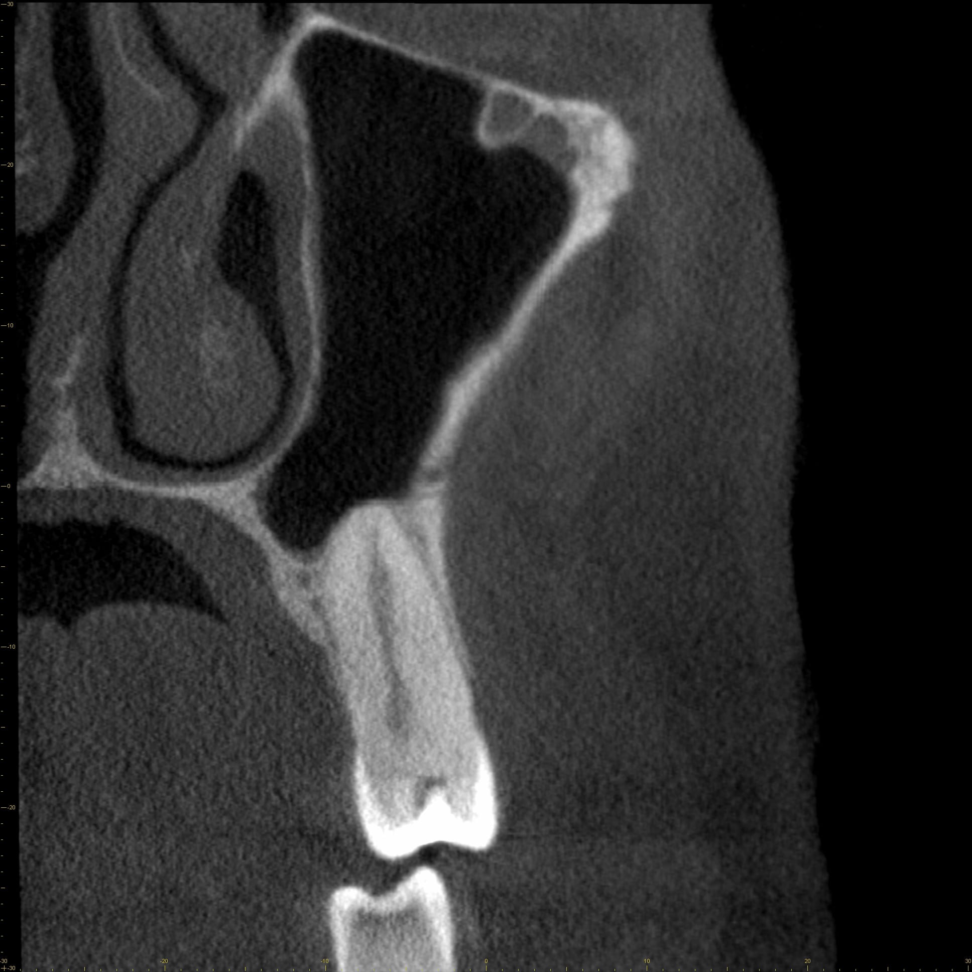
Figure 3B: Coronal CBCT image
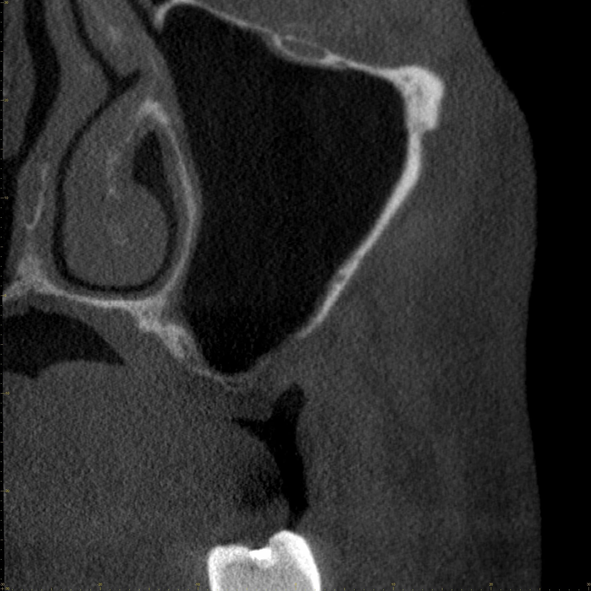
Figure 3C: Coronal CBCT image
Figure 3D: The coronal slice exhibits an enamel pearl in the furcation area of the second molar (*). Based on these findings, a staged sinus floor elevation procedure with implant insertion was planned and discussed with the patient.
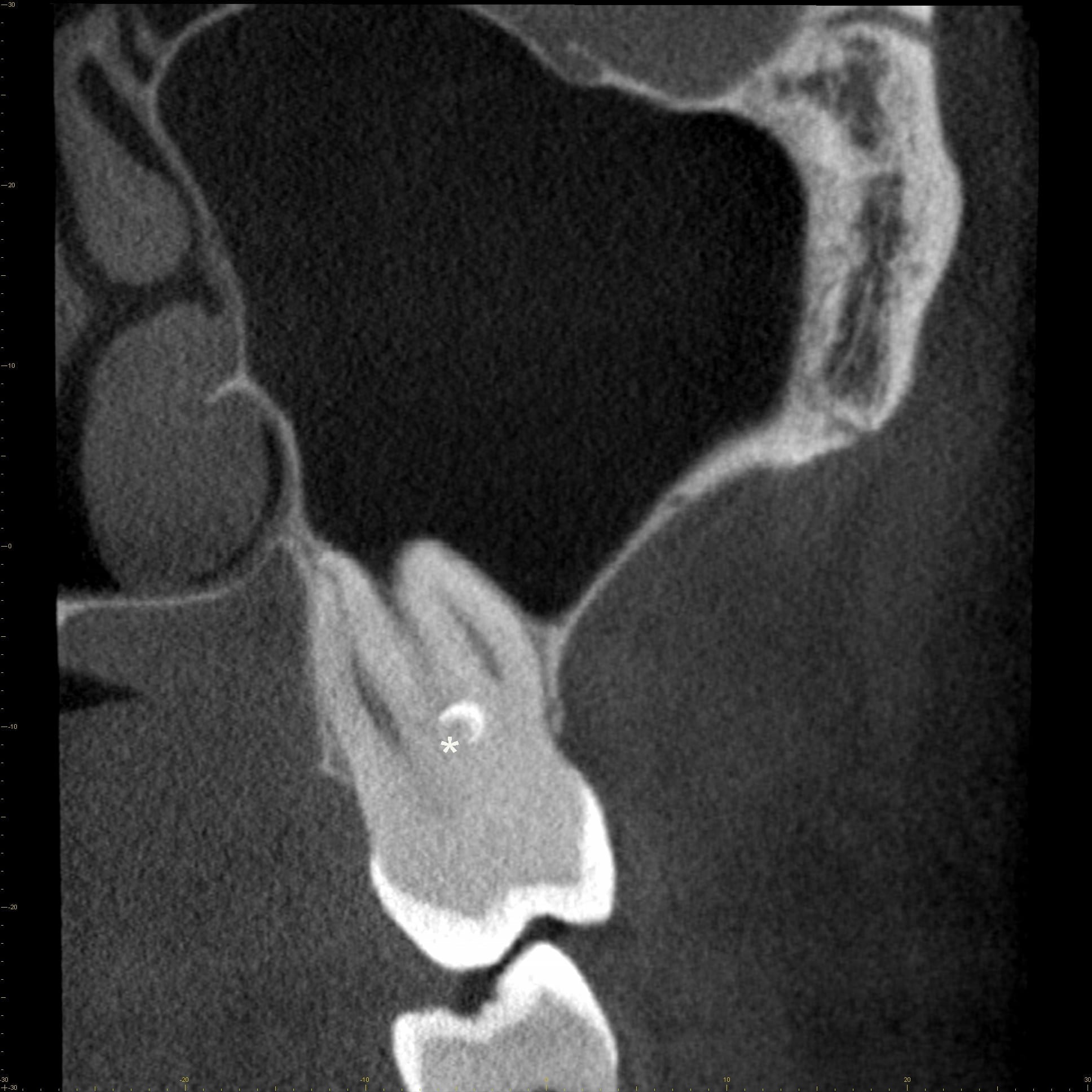
Figure 3D: Coronal CBCT image

Figure 3E: Axial CBCT image


